EQUIPEX — UTEM
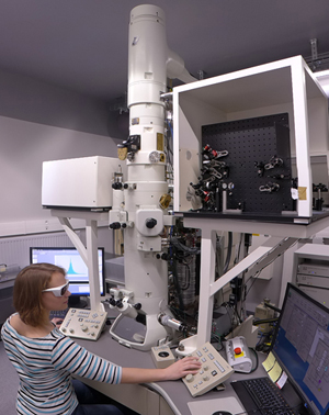
An ultrafast transmission electron microscope has been installed in 2014 at the Institut de Physique et Chimie des Matériaux de Strasbourg (IPCMS). It has been funded in the second round of the national initiative Investissements d’Avenir as an EQUIPEX project. It is one of the world’s first advanced commercial electron microscopes with ultrahigh temporal resolution.
The commercial basis of the microscope has been extended and optimized by us during the first years of operation. Now, the UTEM is operational in the stroboscopic as well as in the single-shot mode. Several scientific projects on applications in nanomaterials science are carried out.
External users in France have access to this microscope through the METSA network. Please contact us if you are interested.
Articles
Some publications of studies with this microscope
K. Bücker, M. Picher, O. Crégut, T. LaGrange, B. W. Reed, S. T. Park, D. J. Masiel, F. Banhart, The electron dynamics in an ultrafast transmission electron microscope with Wehnelt electrode, Ultramicroscopy, 171, 8 (2016)
M. Picher, K. Bücker, T. LaGrange, F. Banhart, Imaging and electron energy-loss spectroscopy using single nanosecond electron pulses, Ultramicroscopy 188, 41 (2018).
K. Sinha, A. Khammari, M. Picher, F. Roulland, N. Viart, T. LaGrange, F. Banhart, Nanosecond electron pulses in the analytical electron microscopy of a fast irreversible chemical reaction, Nature Communications 10, Article 3648 (2019), https://www.nature.com/articles/s41467-019-11669-w
J. Sun, S. K. Sinha, A. Khammari, M. Picher, M. Terrones, F. Banhart, The amorphization of metal particles in graphitic shells under laser pulses, Carbon 161, 495 (2020).
Y. Hu, M. Picher, N. M. Tran, M. Palluel, L. Stoleriu, N. Daro, S. Mornet, C. Enachescu, E. Freysz, F. Banhart, G. Chastanet: Photo-Thermal Switching of Individual Plasmonically Activated Spin Crossover Nanoparticle Imaged by Ultrafast Transmission Electron Microscopy, Advanced Materials 33, 2105586 (2021) https://doi.org/10.1002/adma.202105586
M. Picher, S. K. Sinha, T. LaGrange, F. Banhart, Analytics at the nanometer and nanosecond scales by short electron pulses in an electron microscope, Chem Texts 8, 18 (2022)
Y. Hu, M. Picher, M. Palluel, N. Daro, E. Fresz, L. Stoleriu, C. Enachescu, G. Chastanet, F. Banhart, Laser-driven transient phase oscillations in individual spin-crossover particles, Small 2303701 (2023) https://onlinelibrary.wiley.com/doi/full/10.1002/smll.202303701
H. Mba, M. Picher, N. Daro, M. Marchivie, P. Guionneau, G. Chastanet, F. Banhart, Lattice defects in sub-micrometer spin-crossover crystals studied by electron diffraction, J. Phys. Chem. Lett. 14, 8100 (2023)
——————————————————————————————————————–
Some newspaper and web articles
Dernières Nouvelles Alsace DNA 27/05/2015
Le guide de la R&D Innovation Review
Nature Research Chemistry Community
Institut de Physique – CNRS 2019
Workshop